
Dry Eye
What Causes Dry Eyes?
Dry eyes, medically known as keratoconjunctivitis sicca, occur when the tear ducts do not provide adequate tears to effectively lubricate the eyes. This chronic condition is usually due to decreased tear production, increased evaporation of tears, or an imbalance in the composition of tears.
Decreased tear production
You can develop dry eyes due to decreased tear production naturally as you age. But you can also have this uncomfortable condition due to:
Regularly looking at a computer screen (computer vision)
Chronic diseases, such as diabetes and rheumatoid arthritis.
Laser eye surgery
Lacrimal gland damage
Dry eyes from decreased tear production can even be a side effect of certain medications, particularly decongestants and allergy medications. This is why it is so important to discuss all medications, including over-the-counter varieties, with your optometrist at My Vision Eye Care.
Increased evaporation of tears
Increased tear evaporation means you probably have enough tears, but they don’t effectively lubricate your eyes. This can happen due to:
Wind, pollution, or dry air
Blink less often
Eyelid problems
In some cases, increased tear evaporation can be due to both environmental and physical problems, such as living in a dry climate and having eyelids that turn (ectropion).
Imbalance in the composition of tears
Your tears are made of oil, water, and mucus. But if your sebaceous glands (meibomian glands) become clogged, your tears may not have enough oil to properly lubricate your eyes. In many cases, your optometrist can get to the root of a tear imbalance and correct the underlying problem, so you can start producing healthy tears.
Why should I be concerned about dry eyes?
Dry eye problems don’t just leave you with dry, scratchy, and red eyes, these complications can lead to other serious problems. Untreated dry eyes can:
Increase your risk of eye infections.
Lead to damage to the surface of the eye.
Make it difficult to see and do daily tasks.
The earlier you start treatment for dry eyes, the less likely you are to have these serious complications.
Do you treat urgent or emergency problems related to dry eyes?
Yes, we treat severe red, pink, sore, dry eye conditions and have a dry eye protocol that we follow as it is not a one-size-fits-all solution, but rather a condition that needs to be managed over time.
How are dry eyes treated?
Our doctor will evaluate your eyes to determine what is causing their dryness. Depending on the cause, your dry eye treatment may include:
Medications to reduce inflammation of the eyelids.
Eye drops to minimize corneal inflammation.
Prescription drugs that stimulate tears
Daily eye inserts to lubricate your eyes.
Punctal plugs
Many dry eye patients benefit from point plugs. These inserts go directly to the tear ducts and block the flow of tears from the eye to the nose and sinuses. This forces tears, as well as essential eye drops, to remain in the eyes for longer periods of time.
Before you live another day with dry eyes, see how My Vision can help you. Book your appointment through the online scheduler or call the office.
Ocular Diabetes:
Ocular diabetes is a major problem in eye care in developed countries. It is the leading cause of blindness between the ages of 20 and 64. 25% of diabetics have some form of retinopathy, with 5% of them being a serious form.
Diabetic retinopathy can appear in both juvenile type (type I) and adult (type II) diabetes mellitus.
The risk of developing diabetic retinopathy (DR) is higher the longer the disease has been evolving.
The degree of severity of DR also depends on many other factors: type of diabetes, age of the patient, metabolic control, associated hypertension and / or hypercholesterolemia.
How does hyperglycemia affect eye health?
Through complex mechanisms that are still under investigation, retinal blood circulation undergoes two main disorders in diabetes.
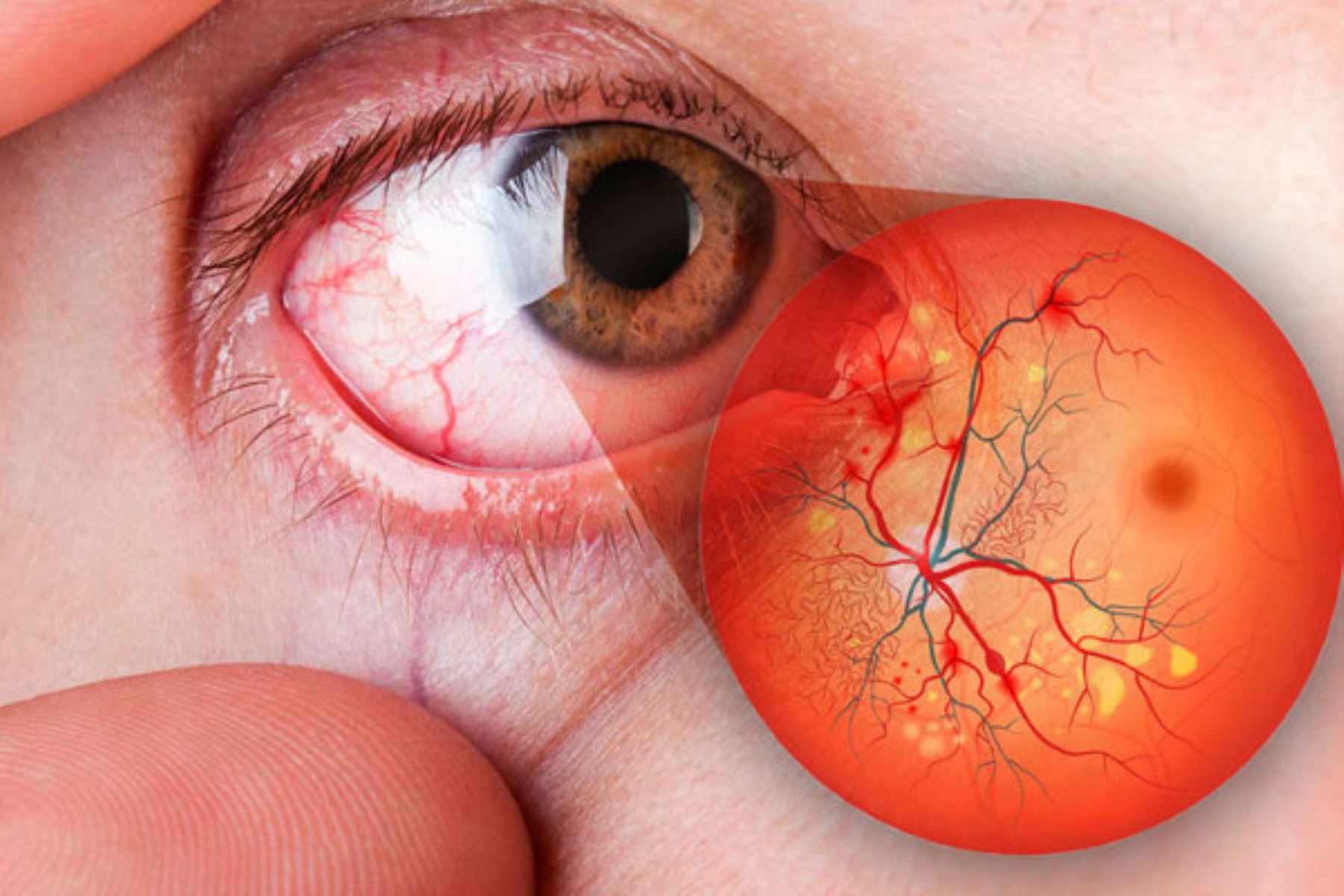
On the one hand, blood flow through the retinal capillaries is reduced (capillary ischemia) with the consequent deficit in the supply of oxygen and nutrients to the retina. The retina is one of the tissues with the highest oxygen consumption in the entire body.
On the other hand, the barrier function of those same capillaries is broken, which in normal situations keeps the retina relatively isolated from the blood. Consequently, the discharge of blood plasma and lipids occurs within the layers of the central retina, called the macula, (macular edema).
Retinal capillary ischemia is the cause of the appearance of neovessels (new vessels that try to provide that deficient oxygen), which lead to two of the most feared complications of diabetic retinopathy: vitreous hemorrhage and tractional retinal detachment. If the ischemia reaches a certain intensity, the neovessels grow distantly over the anterior surface of the iris, which can increase intraocular pressure (neovascular glaucoma)
Forms of ocular diabetes:
waterfall
Diabetic retinopathy
neovascular glaucoma
weakness of the corneal epithelium.
DIABETIC RETINOPATHY: A Growing Concern
Diabetic Retinopathy presents a major obstacle in the field of vision health, especially in developed nations. It’s the primary cause of blindness among people between the ages of 20 and 64, impacting roughly 25% of individuals with diabetes to varying degrees of retinopathy, while 5% of them endure a severe form of the condition.
Diabetic retinopathy can appear in both type I diabetes (juvenile onset) and type II diabetes (adult-onset). The likelihood of developing diabetic retinopathy (DR) rises with the duration of the disease. The severity of DR is affected by multiple factors, such as the type of diabetes, age of patient, the level of metabolic control, and the presence of hypertension and hypercholesterolemia.
Typically, your primary care physician will recommend a visit to a nearby optometrist or ophthalmologist to evaluate your eye health. At Mi Vision in San Antonio, we collaborate with various specialists in situations where advanced stages of this condition require a referral. If the condition is minor, we will assess it at our Balcones Heights office and schedule appropriate follow-ups at intervals of 3, 6, or once a year.
How Does Hyperglycemia or High Glucose Levels Impact
Eye Health?
The effects of hyperglycemia, also known as high blood sugar levels, on the eyes involves complex mechanisms that are still being investigated. In diabetes, two primary disturbances affect the retinal blood circulation. First, there is a decrease in blood flow through the retinal capillaries, resulting in capillary ischemia.
This, in turn, leads to an insufficient delivery of oxygen and nutrients to the retina. The retina is one of the body's most oxygen-dependent tissues.
Secondly, the barrier function of these capillaries becomes compromised, which typically helps isolate the retina from the blood. As a result, blood plasma and lipids are released into the layers of the central retina, particularly the macula, which leads to macular edema.
The development of neovascularization (formation of new vessels attempting to supply the oxygen-deficient areas) is a consequence of retinal capillary ischemia. These neovessels are associated with two severe complications of diabetic retinopathy: vitreous hemorrhage and tractional retinal detachment. In cases of intense ischemia, the neovessels may extend over the anterior surface of the iris, potentially raising intraocular pressure and causing neovascular glaucoma.
When you visit our eye doctor in San Antonio, we will inquire about your medical history and then proceed to dilate your eyes to obtain a clearer view of your retina. Our optometrist will not only examine your retina with the naked eye, but also employ the advanced Optical Coherence Tomography (OCT) Zeiss equipment.
This cutting-edge technology not only captures detailed images of your eyes, but also analyzes the retinal structures and provides a comprehensive evaluation of your retinal condition. We strongly recommend undergoing this eye test annually, regardless of whether you have diabetes or not.
Forms of Ocular Diabetes
1. Cataracts
2. Diabetic retinopathy
3. Neovascular glaucoma
4. Corneal epithelial dystrophies
5. Blurry vision mainly at distance
Given the escalating occurrence of ocular diabetes, it is crucial to adopt a proactive approach in diagnosing, treating, and managing this condition. By intervening early and closely monitoring patients, we can help mitigate the impact of this condition on your vision and overall eye health.
Dr. Cuba and the dedicated team at Mi Vision are committed to assisting you throughout the entire process. We will explain the most suitable treatment options available to you and make referrals to the appropriate local eye care specialists when necessary. The prevalence of diabetes among the population in San Antonio, TX is significant, and we are here to offer our assistance and eye care support to our Alamo City community.
Currently we are offering initial screening assessment with OPTOS technology to assess diabetic retinopathy and other retinal conditions without dilation https://www.optos.com/blog/2017/july/what-is-diabetic-retinopathy/

Cataracts are the formation of a nebula in the lens of the eye, which is behind the iris and the pupil. Cataracts usually occur in both eyes, but sometimes they only affect one. Most cataracts occur as a result of aging, usually sometime after the age of 40.
Cataracts are the most common cause of vision loss worldwide, but they can be treated.
Causes of cataracts
As we age, the proteins that make up the lens can clump together. These clusters are the cataracts and they are what causes this cloudiness. Over time, they can get larger and cloud more of the lens, making vision difficult.
The lens of the eye works like a camera lens, focusing light on the retina for clear vision. It also adjusts the focus of the eye, allowing us to see up close and far.
Most of the lens is made up of water and protein. Proteins are arranged in a specific way that makes the lens transparent and allows light to pass through.
It is not known with certainty why the lens changes as we age, forming cataracts. Researchers around the world have identified factors that may be related to the appearance of cataracts. In addition to aging, risk factors for cataracts include:
UV radiation from sunlight and other sources
Diabetes
Hypertension
Obesity
Smoking habit
Long-term use of corticosteroids
Using statins to lower cholesterol
Previous eye injury or inflammation
Previous eye surgery
Hormone replacement therapy
Significant alcohol consumption
High myopia
Family background
One current theory is that oxidative changes in the human lens may be the cause of cataracts. Studies have shown that fruits and vegetables high in antioxidants can help prevent some types of cataracts.
Cataract treatment
When symptoms start to appear, you may be able to improve your vision for a time by using:
New glasses.
Powerful bifocals.
Augmented glasses.
Adequate lighting or other visual aids.
Other treatment options for cataracts that have progressed may include cataract surgery.
If cataracts begin to affect your quality of life, your ophthalmologist may suggest surgery. This is generally considered an effective, low-risk way to restore vision.
Poor vision appears to many people to be an inescapable consequence of aging, but cataract surgery is a simple and relatively painless procedure to restore vision.
Cataract surgery is very successful in restoring vision. It is the most commonly performed type of surgery in the United States, and more than 2 million Americans undergo it each year, according to the Prevent Blindness organization.

DMRE causes you to lose central vision. He cannot see the fine details, not near or far. However, peripheral (side) vision works normally. For example, imagine you are looking at a clock with hands. If you have AMD, you may be able to see the numbers on the clock but not the hands.
AMD is very common. It is one of the main causes of vision loss in people over 50 years of age.
Two types of AMD
Dry AMD
This type of degeneration is quite common. About 80% (8 out of 10) of people with AMD have the dry type of degeneration. Dry AMD occurs when parts of the macula become thinner with age, causing a clump of proteins called drusen. This causes you to lose central vision slowly. There is no treatment yet for dry AMD.
When you have AMD, dark areas may appear in your central vision.
DMRE WET
This type is less common but much more serious. Wet DMRE occurs when abnormal blood vessels grow under the retina. These vessels can leak blood or other fluids, causing scarring of the macula. Vision is lost faster with wet DMRE than with dry DMRE.
Many people do not realize they have DMRE until their vision has become very blurry. This is why it is so important to visit an ophthalmologist regularly. Your ophthalmologist or ophthalmologist can look for early signs of DMRE before you have vision problems.
You are more likely to have DMRE if:
Having heart disease is also a risk factor for DMRE, in addition to having high cholesterol levels. Caucasians (white people) are also at higher risk of developing DMRE.
DMRE gradually causes vision changes. You may not notice these changes when they occur. But you need to identify them as soon as possible. Treating them early can help slow or stop vision loss.
DMRE gradually causes vision changes. You may not notice these changes when they occur. But you need to identify them as soon as possible. Treating them early can help slow or stop vision loss:
This is what the Amsler grid might look like when its areas have blurred and wavy lines.
Call your ophthalmologist right away if you notice any lines or parts of the grid look wavy, blurry, or dull.
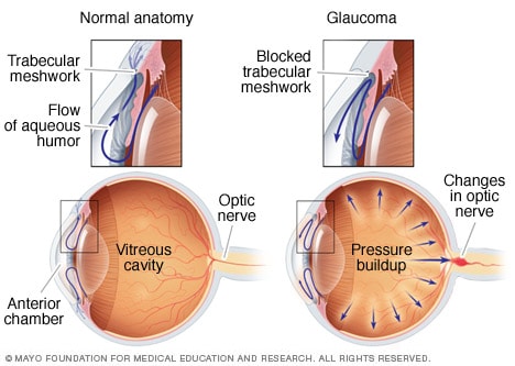
Glaucoma is the leading cause of blindness in people over 60 years of age. Often times, blindness due to glaucoma can be prevented if it is treated early.
The eye constantly produces aqueous humor. As new aqueous humor flows into the eye, the same amount must be drained. The fluid drains through an area called the drainage angle. This process keeps the pressure in the eye (called intraocular pressure or IOP) stable. However, if the drain angle is not working properly, fluid builds up. The pressure inside the eye increases and this damages the optic nerve.
If the drainage angle is blocked, fluid cannot leave the eye, causing an increase in pressure.
The optic nerve is made up of more than a million small nerve fibers. It is similar to an electric cable made up of many small wires. When these nerve fibers die, blind spots develop in vision. You may not notice these points
Glaucoma symptoms
Symptoms of open-angle glaucoma
Open-angle glaucoma has no warning signs or obvious symptoms during the early stages. As the disease progresses, blind spots develop in peripheral (side) vision.
Most people with open-angle glaucoma do not notice any change in their vision until the damage is quite severe. That is why glaucoma is called the “silent thief of vision.” Keeping a plan for regular eye exams can help your ophthalmologist discover disease before vision is lost. Your ophthalmologist can tell you how often you should be seen.
Symptoms of angle-closure glaucoma
People at risk of developing angle-closure glaucoma usually show no symptoms before an attack. Some of the initial symptoms of an attack may include blurred vision, halos of light, mild headaches, or pain in the eye. People with these symptoms should be seen by an ophthalmologist as soon as possible. An attack of angle-closure glaucoma includes the following symptoms:
severe pain in the eye or forehead
eye redness
decreased vision or blurred vision
vision of rainbows or halos of light
headache
nausea
threw up
blind until most of the optic nerve fibers have died. If all the fibers die, you will go blind.